Dense Breasts? No Compression Imaging Can Help
Posted by Pabitra Giri
Filed in Health 232 views
The Letter That Sends Women Scrambling for Answers
In recent years, many US states have required that women be notified when their mammogram shows dense breast tissue. For a lot of women, that letter arrives without much context — just a statement that their breast tissue is dense and that this may affect the sensitivity of their mammogram, and a suggestion to talk to their doctor.
The conversation that follows is often frustrating. Dense breast tissue is common, affects about 40 percent of women in the US, and genuinely complicates breast cancer detection on standard mammography. But the options available for follow-up imaging have historically been limited in clarity, comfort, and accessibility.
No compression breast imaging represents a meaningful shift in what those follow-up options look like. For women who have been told they have dense breasts and are trying to understand what that actually means for their health and their imaging choices, this blog is written specifically for you.
What Dense Breast Tissue Means and Why It Matters
Breast density is a measure of how much fibroglandular tissue a breast contains relative to fatty tissue. On a mammogram, fatty tissue appears dark, while fibroglandular tissue and tumors both appear white or light gray. When there's a lot of fibroglandular tissue — when breasts are dense — the white-on-white masking effect can make it genuinely difficult to distinguish abnormal findings from normal tissue.
This creates two distinct problems. The first is detection sensitivity: studies consistently show that mammogram sensitivity is lower in women with dense breast tissue, meaning some cancers that would be visible in fatty breast tissue are missed in dense tissue. The second is specificity: the overlapping white tissue creates findings that look potentially concerning but turn out to be normal tissue seen at an ambiguous angle, which drives callback rates and subsequent imaging for findings that aren't pathological.
Dense breast tissue is also an independent risk factor for breast cancer — women with very dense breasts have approximately four to six times the breast cancer risk of women with mostly fatty tissue. So the population most affected by mammogram's limitations is also the population with elevated risk. That's a meaningful gap in the current standard of care.
Why Standard Alternatives Have Limitations Too
When women with dense breasts seek additional imaging, they're typically offered ultrasound or MRI. Both have genuine utility, but both also have real limitations in this context.
Ultrasound is widely available and doesn't involve compression, but it's highly operator-dependent, has a significant false positive rate, and doesn't produce the kind of precise anatomical mapping that guides clinical decision-making with confidence. It's useful for characterizing specific known findings, but it's limited as a comprehensive imaging approach for dense breast tissue.
Breast MRI is the most sensitive imaging modality available for breast cancer detection, but it requires contrast injection, takes 30 to 45 minutes, is expensive, and is typically reserved for high-risk patients — BRCA mutation carriers, women with a strong family history, those with prior chest radiation. For the large population of women with dense breasts who are at intermediate rather than high risk, MRI is often not clinically justified or practically accessible.
That's the gap that 3d no compression breast imaging fills — an imaging approach that's more informative than mammography in dense tissue, more comfortable than any compression-based modality, more accessible and faster than MRI, and validated at the highest level of FDA clearance for diagnostic breast imaging.
The Technology Behind the Difference
The Koning Vera system produces true three-dimensional cone-beam CT images of the breast. Understanding why this matters requires understanding what makes 3D imaging different from what's been available before.
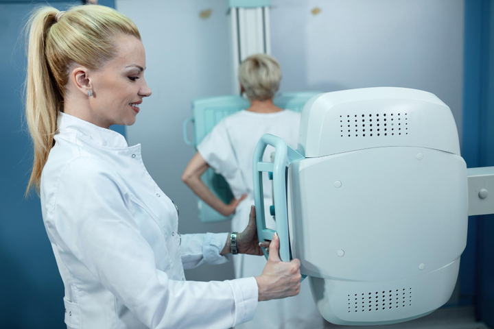
Standard mammography produces a 2D projection — a flat image created by compressing all the breast tissue into a single plane and passing X-rays through it. Digital breast tomosynthesis, often marketed as 3D mammography, creates a series of 2D slices through compressed tissue — it's an improvement over standard mammography, but it still requires compression and still generates slice-by-slice 2D images rather than true isotropic 3D data.
3D breast CT is genuinely different. The Koning Vera system rotates 360 degrees around the breast as it hangs naturally below the patient, capturing data from every angle simultaneously. The resulting dataset is fully three-dimensional and isotropic — meaning the resolution is identical in every plane, and a radiologist can examine the breast tissue from any orientation, any slice angle, with the same image quality throughout.
No compression is required because the 3D data acquisition doesn't need flat tissue — it captures the breast as it naturally exists, without distortion, without the masking effect of overlapping tissue compressed into a single plane. The scan takes about seven seconds per breast. The total bilateral exam takes under five minutes.
Radiation exposure is comparable to a standard 2D mammogram and adheres fully to MQSA limits — approximately 0.7 mSv for a non-contrast scan, which is about 75 percent less than the average person's annual background radiation exposure.
What No Compression Breast Imaging Looks Like in Practice
Gnosis for Her brings the Koning Vera technology to Southern California communities through a mobile care unit — a thoughtfully designed space that prioritizes the patient experience from the moment a woman arrives. There are no large hospital corridors, no waiting room anxiety, no cold clinical environment.
The scan itself is straightforward. The patient lies face down on a specialized table. Each breast rests naturally in an opening below the table, and the system completes its 360-degree rotation around the breast in approximately seven seconds. No plates. No compression. No instructions to hold still while something uncomfortable happens.
The images produced are read by a board-certified radiologist. Results are delivered securely to the patient and their physician, typically within 72 hours when prior imaging is available. The entire experience — from arrival to departure — is designed around what a woman with a real breast health concern actually needs: clear information, delivered with care, without adding unnecessary barriers or discomfort.
Specific Situations Where This Imaging Is Most Valuable
Gnosis for Her is transparent about where 3D Breast CT sits in the clinical picture — it's diagnostic imaging designed to complement mammography, not a general screening replacement. The women who benefit most are those who need more than a standard screening can provide.
Women who have received a BI-RADS 3 assessment — probably benign, follow up in six months — can use 3D Breast CT to get a more definitive answer about the finding rather than living with uncertainty through another half-year waiting period. Women with palpable lumps, nipple changes, focal pain, or other breast symptoms benefit from the comprehensive 3D view that guides clinical decision-making with confidence. Women who have implants and have been told their mammogram results are limited by the presence of the implants can receive clear imaging of the breast tissue without the distortion that compression creates.
And women who have simply been postponing imaging because they can't tolerate the discomfort of compression — a more common situation than most clinical settings acknowledge openly — finally have an option that removes that barrier entirely.
The Accessibility Mission Behind the Technology
What sets Gnosis for Her apart from a standard imaging center isn't only the technology — it's the explicit commitment to health equity. Barriers like transportation, lack of a primary care provider, and out-of-pocket costs prevent many women from accessing the breast imaging they need. The mobile clinic model directly addresses the transportation barrier by bringing the technology to communities rather than requiring communities to travel to it.
The partnership with Karis Healthcare addresses the provider barrier, offering a brief telehealth evaluation for women who need the required physician order but don't have easy access to a primary care provider. Transparent self-pay pricing at $499 — with FSA/HSA reimbursement eligibility and planned insurance acceptance — reflects a genuine effort to make advanced breast imaging accessible to women across income levels, not only to those with comprehensive commercial insurance.